
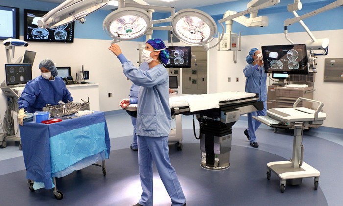
The new T-Suite at M Health Fairview University of Minnesota Medical Center, named for its shape, is a combination of clinical technology and research that is the first of its kind. The four-room surgical suite is home to a powerful, mobile magnetic resonance imaging (MRI) scanner. The scanner travels between the three operating theaters within the suite, where experts will use it to view real-time images of the brain during surgery.
This capabilities of this space will help University of Minnesota Physicians (M Physicians) neurosurgeons, radiologists and others redefine treatment options and come together to provide world-class surgical care.
“The space will fundamentally change how we can treat our patients. For example, if you have a stroke, today you will be imaged in an MRI located far away from where your actual procedure will occur. After the MRI, you'll be transported from that space to the procedure room. With this particular arrangement, you can be imaged in the MRI and receive the treatment that you need within minutes."
Clark Chen, MD, PhD, M Physicians neurosurgeon, Lyle French Chair in Neurosurgery and the head of the University of Minnesota Medical School Department of Neurosurgery.
“We have been well-known for our international research in MRI and other fields. But bringing that to the cutting-edge of clinical delivery has always been a bit difficult because there are many more considerations when it comes to safety and sterility when you have operating rooms. So, we are very excited about it, because now we can bring some of those things directly to our patients that we've been working on for a decade or two,” said Alexander McKinney, MD, M Physicians radiologist.
One of the rooms can be used for laser ablation, which cannot be performed in a traditional operating room setting. Another can be used for endovascular (catheter-based) procedures like embolization and stroke treatment. Through this multi-room setting, a person undergoing a complex, multi-stage surgery can move for each stage of their procedure, often only needing to undergo anesthesia once. This allows their treatment to be completed over the span of one day as opposed to multiple.
The MRI scanner, which travels on a ceiling-mounted rail system between and into the operating rooms, provides diagnostic-quality intraoperative images. The real-time imaging provided by the surgical suite can also improve outcomes for people with other neurological conditions, including stroke and Parkinson’s disease. Instantaneous feedback helps physicians understand how neural connectivity changes in the brain as surgery progresses, allowing specialists a better opportunity to tailor surgical treatment to the patient.
The T-Suite also establishes the facility as a destination for other physicians and researchers to visit and enhance their knowledge. Dr. Chen said, “One of the most exciting aspects of this arrangement is that we will have an observer deck where people from all over the world can come and study how we perform surgeries and how we advance the understanding of the human brain.”
Patients are the obvious benefactor of the suite’s capabilities, but Drs. Chen, McKinney and others are just as eager to utilize the groundbreaking space.
“When I saw [the T-suite], I was very excited because we're finally bringing to our patients what we've theoretically wanted to do for years—bringing cutting-edge research into the clinical realm,” said Dr. McKinney, who is also the vice-chair of Research and Informatics and Neuroradiology director in the Radiology Department at the U of M Medical School.
“I believe that this suite will be the canvas on which we paint the neurosurgeries of tomorrow,” Chen said. “Seeing this come true is like seeing the sunrise on top of the Himalayas. It’s that extraordinary feeling that you can't quite explain.”