
Peter Hilger, M.D., Maurice M. Khosh, M.D., Gary Nishioka. M.D., D.M.D., and Wayne F. I.arrabee, M.D.
Minneapolis, Minn., unit Seattle. Wash.
The Mustarde technique is one of the most commonly used methods for creation of the antihelical fold in the prominent ear deformity. One part of this procedure that can be difficult to perform is precise tightening of the cartilage conforming sutures. Because the surgeon’s attention is directed laterally to achieve the desired antihelical shape, tightening the sutures medially generally is done without direct exposure. This maneuver is awkward and creates a risk of under correction by tying the knots too loosely because of soft-tissue entrapment and the tendency for the cartilage to unfold. Our modification facilitates placement of Mustardé. These contouring sutures fix the antihelical fold in a stable position, which allows carefree manipulation of the auricle while permanent sutures are placed through the postaurircu1ar incision and tightened under direct vision.
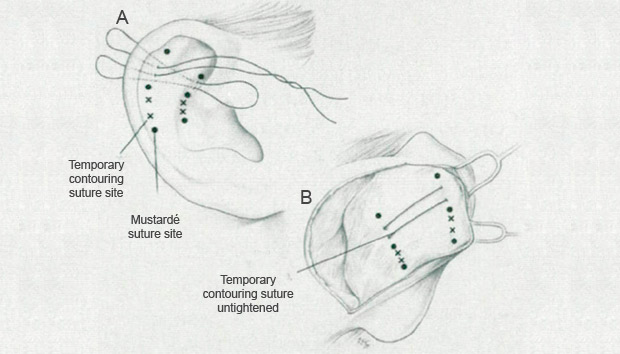
Fig. 1. Placement of the temporary contouring sutures.
TECHNIQUE
Typically, three sutures sites are marked. The standard postauricular incision is followed by a supra-perichondrial dissection toward the helical rim to expose the dye marks. For the temporary contouring sutures. 3-0 polypropylene sutures, double-armed with large Keith needles, are used. In general, two horizontal mattress sutures placed between the planned Mustardé sutures are sufficient.
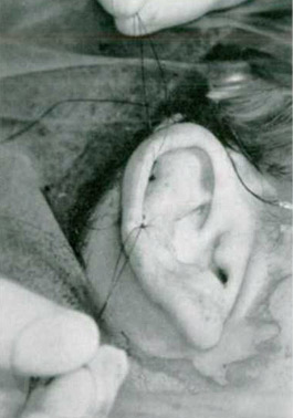
FlG. 2. Tightening and tying of the temporary sutures.
The first contouring suture is placed between the upper and the middle dye marks. The Keith needles are passed from the lateral aspect near the conchal bowl and withdrawn through the wound. Each needle is then passed through the wound, close to the helical rim, and withdrawn through the lateral auricular skin (Fig. 1). This completes the first mattress suture. The next suture is similarly placed between the middle and lower dye marks.
Once both sutures are placed, they are tightened until the desired antihelical fold is achieved and then tied (Fig. 2). The newly shaped auricle can now be manipulated in any manner to provide maximal exposure of the postauricular wound. Next, permanent Mustardé sutures are placed and tied under direct vision (Fig. 3). The permanent sutures should be lightened just to the point that tension is taken off the temporary contouring sutures. This will ensure the desired antihelix shape after the contouring sutures are removed. Additional maneuvers such as conchal bowl reduction or set-back can be performed at this time. The contralateral ear is addressed subsequently.
SUMMARY
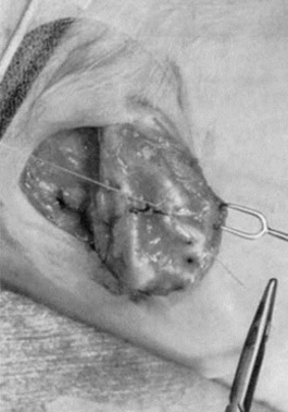
FlG. 3. Placement of Mustardé sutures under direct vision.
Temporary contouring sutures simplify precise suture placement in the Mustardé otoplasty technique. This modification allows the surgeon to fold the ear forward, expose the postauricular incision, and place and tighten the permanent sutures under direct vision. Contouring sutures eliminate the risk of antihelical fold change by maintaining the cartilage in a predetermined position while the ear is manipulated. We have found this maneuver to be effective, safe, and easy to perform.
Wayne F. Larrabee, M.D. Suite 280 600 Broadway Seattle, Wash. 98122
REFERENCES
- Bull. T. R.. and Mustardé J. C. Mustardé technique in otoplasty. Facial Plastic Surg. 2(2): 101, 1985.
- Mustardé, J. C Correction of prominent ears using simple mattress sutures. Br. J. Plast. Surg. 16: 170. 1963
- Tardy, M. E. Jr., Tenta, L. T., and Pastorek, N.J. Mattress suture otoplasty: Indications and limitations. Laryngoscope 79: 961, 1960.
From the Department of Otolaryngology – Head & Neck Surgery, University of Minnesota, Minneapolis, and the Department of Otolaryngology-Head & Neck Surgery, University of Washington, Seatle. Dr. Nishioka is in private practice in Seattle. Received for publication December 18, 1996; revised March 14, 1997.